Cutaneous Lymphoma and Tumormicroenvironment
Prof. Dr. med. Christina Mitteldorf
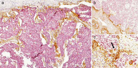
- Cutaneous lymphomas: Improvement of diagnostic methods for early tumor detection and lymphoma classification. Systematic and standardized evaluation of checkpoint and targeted molecules.
- Cutaneous Pseudolymphomas: We investigate human samples by histology, immunohistochemistry and molecular diagnostics to find discriminating factors from cutaneous lymphomas.
- Tumormicroenvironment and PD1 and PD-L1 expression in cutaneous neoplasms: We use immunohistochemical and immunofluorescence multi stains to characterize the composition of the TME and to image relations between different cell types. One intention is to localize targeted molecules (e.g. PD-L1) on different cell types.
- Digital image analysis: We use digital image analysis to evaluate the expression of surface proteins.
- Development of a digital learning platform for dermatopathology
Cutaneous lymphomas:
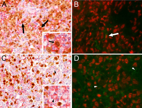
Tumormicroenvironment and PD1 and PD-L1 expression in cutaneous neoplasms:
Team
- Viktoria Amon
- Dr. Johanna Hoffmann
- Noman Langer
- Franziska Kampa
- Aleksandra Kulberg
- Nikola Rapsch
Contact
Oberärztin
Kontaktinformationen
- Telefon: +49 551 3968082
- E-Mail-Adresse: christina.mitteldorf(at)med.uni-goettingen.de
- Oberärztin / Fachärztin für Dermatologie und Venerologie, Medikamentöse Tumortherapie, Dermatopathologie
- International Board Certification in Dermatopathology
- Oberärztliche Leitung der Dermatopathologie und der Lymphomsprechstunde
- Pubmed-Link